Up to 50% off NYC founding offers.
Learn More
Myo Brentwood, 90049
U.S.
English
English (Canada)
my account
STORE
Treatments
Shop Treatment Tools
Locations
Shop Apparel
Back to Main Site
Our Story
Contact Us
My Account
Book Now
Book Now
Myo Brentwood, 90049
Book Now
THE MYO BLOG
FutureProof Journal
All
Education
News
Culture
Our Founder’s Interview
Discover Myodetox’s founding story—Vinh Pham and Scott Marcaccio share their journey, vision, and ambitious future in physical therapy.
All
1
Min Read
Read article
Body IQ: Making Sense of Your Body
Education
3
Min Read
2025 Myodetox Emerging Leaders Scholarship Program
News
2
Min Read
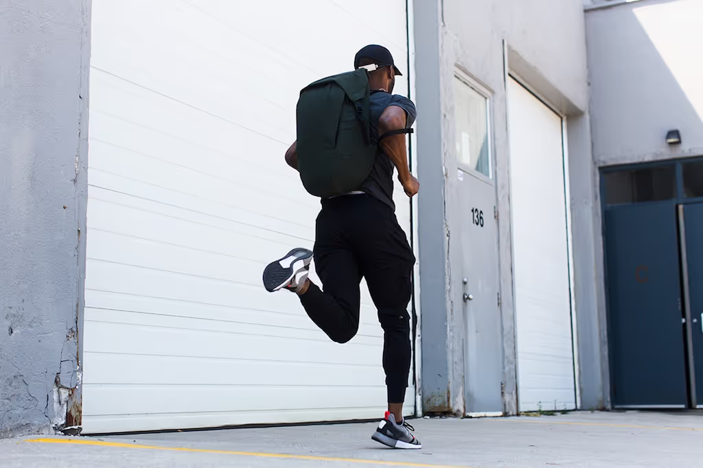
Dr. Kevin’s Top 3 Drills to FutureProof Your Run
Education
2
Min Read
This Running Workout Is Designed To Build Endurance
Education
3
Min Read
Avoid Hip Pain By Adding Three Exercises To Your Daily Routine
Education
2
Min Read
Increase Your Mid-Back Mobility With These Three Exercises
Education
2
Min Read
Breast Cancer + Pelvic Health
Education
2
Min Read
Meet Your Mississauga Team
Culture
1
Min Read
Osteoarthritis 101
Education
3
Min Read
Protect Your Hamstrings
Education
3
Min Read
What Is A Rotator Cuff Tear?
Education
3
Min Read
The Key To Managing Sciatica
Education
2
Min Read
How to fix IT Band Syndrome
Education
2
Min Read
Strengthen Your Ankle Tendons
Education
2
Min Read
What is Pelvic Floor Physical Therapy?
Education
3
Min Read
How to come back from runner’s knee
Education
3
Min Read
What Is Frozen Shoulder?
Education
2
Min Read
4 Moves Every Cyclist Needs
Education
1
Min Read
5 Exercises For Moms
Education
2
Min Read
Low Back Pain 101
Education
4
Min Read
What are Tension-Type Headaches?
Education
2
Min Read
5-minute Exercise Routine for Office Workers
Education
2
Min Read
What’s been happening?
Culture
1
Min Read
3 Types of Shoulder Instability
Education
1
Min Read
3 Ways to Prevent Shin Splints
Education
3
Min Read
Test Your Hip Mobility
Education
3
Min Read
How to Treat a Pinched Nerve in Your Neck
Education
2
Min Read
Our Founder’s Interview
Culture
1
Min Read
Myo Run Club
Culture
1
Min Read
Hypermobility Explained: More Than Just Party Tricks?
Education
2
Min Read
Joint Swelling – Is it Effusion or Edema?
Education
1
Min Read
Emerging Leaders Scholarship Program
News
2
Min Read
Prevent Text Neck Pain With These Three Exercises
Education
3
Min Read
Relieve Low Back Pain With These Work From Home Exercises
Education
2
Min Read
A Statement from our Founders
Culture
2
Min Read
Kevin Marryshow on Developing His Team
News
2
Min Read
Jessie Wong Wanted To Be More Than A Therapist
News
1
Min Read
Try Bending Your Knees With Knee Tendonitis
Education
2
Min Read
All You Need To Know About Knee Bursitis
Education
3
Min Read
What Is A Shin Splint?
Education
2
Min Read
The Gluteus Medius Stretch Is Key To A Stronger Butt
Education
3
Min Read
DIY: Avoid Muscle Aches During Your Flight
Education
3
Min Read
Don’t Let An ACL Injury Be Your Downfall
Education
6
Min Read
What Is Whiplash?
Education
3
Min Read
Shoulder Impingement Shouldn’t Impede On Your Daily Tasks
Education
3
Min Read
Beginner & Advanced Rotator Cuff Exercises For Shoulder Pain
Education
5
Min Read
Plantar Fasciitis Stretches To Heal Those Heels
Education
4
Min Read
How To Maintain Good Posture While Driving
Education
3
Min Read
Your 4 Step Guide To Fixing Neck Pain Headache
Education
4
Min Read
Top 5 Muscle Pain Relief Stretches You Can Do Anywhere
Education
6
Min Read
3 Exercises To Help With Sciatica Pain Relief
Education
3
Min Read
Life Hacks: Self Myofascial Release Tools To Use At Home
Education
3
Min Read
5 Things You Can Do To Sleep Better At Night
Education
3
Min Read
Five Commute Exercises To Do For Neck Pain and Lower Back Pain
Education
3
Min Read
How To Bar Hop Without Lower Back Pain
Education
3
Min Read
How To Avoid Carpal Tunnel
Education
3
Min Read
How To Spoon Without Shoulder Pain
Education
2
Min Read
How To Fix Your Tight Hips
Education
2
Min Read
How To Carry Your Bag And Avoid Shoulder Pain
Education
3
Min Read
A Weak Butt Causes Hip Pain
Education
3
Min Read
Four Text Neck Exercises To Avoid Text Neck Syndrome
Education
2
Min Read
How To Netflix And Chill Without The Back Pain
Education
3
Min Read
How To Properly Sit At Your Desk
Education
2
Min Read
It’s Time You Fix Your Arch Without Orthotics
Education
1
Min Read
Your Knee Pain, Lower Back and Hip Pain is Because of Your Feet
Education
2
Min Read
Forward Head Posture Is Affecting Your Brain
Education
1
Min Read
This Is Why You Don’t Have Good Posture
Education
1
Min Read
Why Wait For The Pain?
Education
3
Min Read
Your achilles tendon can either make or break you
Education
2
Min Read
This is the only workout you need to know this offseason
Education
4
Min Read
Revolution in Motion
Culture
6
Min Read
Trust Your Struggle
Culture
3
Min Read
See More Articles
It’s time for a 1:1 with a body expert.
book a session

.jpg)